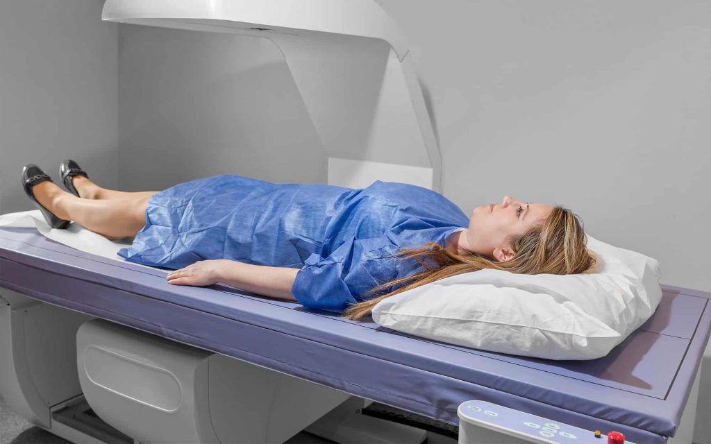
Radiation exposure from bone density scanning represents minimal health risks due to the extremely low X-ray doses required for accurate bone measurements. DXA Scan radiation levels typically range from 1-10 microsieverts per examination, significantly lower than natural background radiation exposure or standard medical imaging procedures. These minimal exposure levels make bone density testing safe for regular monitoring while providing essential diagnostic information for osteoporosis management.
Radiation exposure measurements
DXA examinations deliver approximately 1-6 microsieverts of radiation exposure, equivalent to 1-2 days of natural background radiation from environmental sources. Spine and hip scans typically require the lowest doses within this range, while whole-body examinations may approach the higher end due to increased scanning area. Effective dose measurements account for radiation sensitivity differences among body tissues, providing standardized comparison metrics for various imaging procedures. DXA Scan rank among the lowest radiation exposure medical procedures, comparable to airline flights or natural environmental exposure.
Regional variations in radiation dose occur based on body thickness, scanning technique, and equipment calibration. Technologists optimize scanning parameters to achieve diagnostic quality images while minimizing radiation exposure through proper technique and equipment settings. Modern DXA systems incorporate dose-reduction technologies that maintain image quality while reducing patient radiation exposure compared to older scanning equipment.
Safety comparison standards
DXA radiation exposure represents approximately 1% of annual natural background radiation exposure, demonstrating the minimal nature of these diagnostic procedures. Comparison with other standard medical imaging helps contextualize DXA safety profiles for patient education and informed consent.
- Chest X-rays deliver 5-10 times more radiation than complete DXA examinations
- CT scans expose patients to 100-1000 times more radiation than bone density tests
- Mammography screening involves 10-15 times higher radiation doses than DXA scans
- Annual background radiation exposure exceeds 100 DXA scan equivalents
- Airplane flights expose passengers to similar radiation levels as DXA examinations
International radiation protection organizations classify DXA scans as minimal-risk procedures suitable for routine monitoring without cumulative dose concerns for most patients.
Patient protection protocols
Radiation safety protocols ensure minimal exposure while maintaining diagnostic image quality through proper equipment operation and patient positioning. Lead shielding protects radiosensitive organs outside the scanning area when clinically appropriate and technically feasible. Pregnancy screening prevents inadvertent fetal radiation exposure through comprehensive patient history review and appropriate pregnancy testing when indicated. Alternative diagnostic methods are recommended for pregnant patients requiring bone density assessment. Pediatric protocols incorporate modified scanning parameters that reduce radiation dose while maintaining diagnostic accuracy for younger patients with different tissue characteristics—age-appropriate exposure limits guide pediatric DXA examination protocols. Patient education regarding radiation exposure helps alleviate concerns while providing realistic risk perspectives based on scientific evidence and regulatory guidelines.
Dose monitoring systems
Built-in dosimetry systems track radiation output and patient exposure levels throughout DXA examinations, ensuring consistent safety standards and regulatory compliance. Automatic exposure control optimizes imaging parameters while minimizing radiation dose for each patient.
- Real-time dose monitoring displays radiation levels during scanning procedures
- Quality assurance programs verify dose accuracy and equipment calibration regularly
- Patient dose recording maintains exposure documentation for medical records
- Regulatory compliance monitoring ensures adherence to radiation safety standards
- Equipment maintenance schedules prevent dose drift and maintain safety parameters
Technologist training emphasizes radiation safety principles and dose optimization techniques that protect patients while maintaining diagnostic image quality standards. DXA scan radiation levels represent minimal health risks due to the extremely low X-ray doses required for bone density measurements. These exposure levels fall well below safety thresholds established by international radiation protection organizations while providing essential diagnostic information for osteoporosis prevention and treatment.